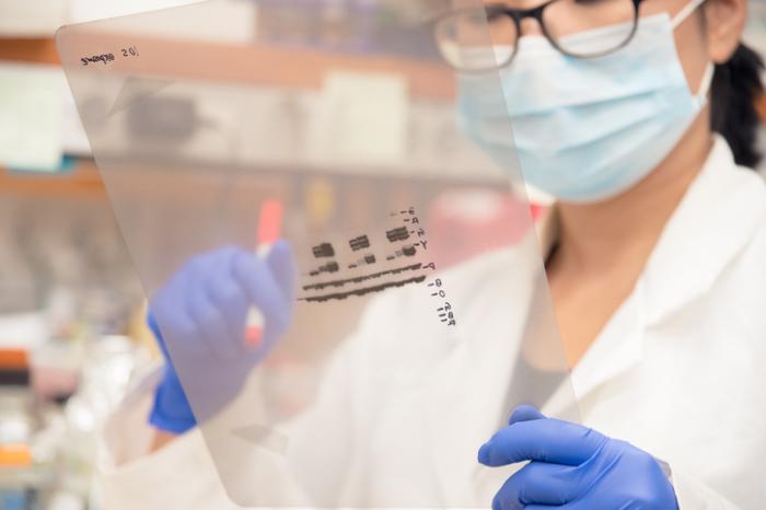
HIV positivity can only be confirmed by the presence of the proteins gp160, gp120, p24, and p31. The positive control row contains proteins from patient sera as well as HIV proteins. HIV is composed of a number of different proteins, which the Western Blot tests for. HIV infected cells are lysed and blotted onto a membrane in the same process.
#WESTERN BLOT VS ELISA HIV FREE#
After the excess second antibody is washed free of the blot, a substrate is added that will precipitate upon reaction with the conjugate resulting in a visible band where the primary antibody bound to the protein. In order to detect the antibodies that have bound, a second antibody is added. If there are any antibodies present which are directed against one or more of the blotted antigens, those antibodies will bind to the proteins and the other antibodies will be washed away. To detect the antigen blotted on the membrane, a primary antibody is added and incubated with the membrane. All sites on the membrane that don’t contain the blotted protein from the gel can then be non-specifically “blocked” so that antibody will not bind to them, causing a false-positive result. The proteins are placed into a gel and an electric current is applied, causing the separated proteins to migrate through the gel and separate according to size and charge. After proteins are separated, they are transferred to a solid membrane through electrophoresis for Western Blot analysis. It allows scientists to visualize antibodies directed against each viral protein. While ELISA measures antibody to whole virus and gives a positive, negative, or indeterminate result, Western Blotting is a more specific test to measure antibodies for HIV. If the test is positive, the patient will be retested, and if the ELISA retest is positive, the patient will then be retested by western blot analysis. A positive result would be 0.500 or higher, 0.300-0.499 need to be retested, and values below 0.300 are considered negative. The results of ELISA are expressed as numbers at a density of 450nm. This can occur during the window between infection and an antibody response to the virus. As HIV buds from the surface of the host cell, it incorporates some of the host cell HLA into its envelope. One reason might be that some people may possess antibodies directed against human leukocyte antigens, which are present on the host cells use to propagate HIV. It is possible that an HIV- person has antibodies which may give a false positive result in the HIV ELISA. Chromogen or substrate will change color when interacting with the enzyme attached to the second antibody. Anti-human immunoglobulin coupled to an enzyme is the second antibody that binds to the human antibodies.

If the patient is HIV+, then the serum will contain antibodies to HIV and will bind to the antigens on the plate. Partially purified, inactivated HIV antigens are pre-coated onto the ELISA plate. It is performed in a plastic plate that contains 96 wells. It is the most basic test to determine if someone is positive for a selected pathogen. ELISA, sometimes called an HIV enzyme immunoassay (EIA) is a tool used to detect HIV based on antibody-antibody interaction.


 0 kommentar(er)
